Sunday, November 18, 2012

Week 5 SIDE EXPERIMENT with (ETOH)


ETOH  EXPERIMENT
 I had been thinking that it would be great, if I could somehow put a paralizing agent into the water, so I could observe some of organisms that moved to quickly to view otherwise. I went to Dr. McFarland, and asked him what he thought about me adding a little, Ethyl Alcohol to the water to kill the organisms so I could view the ones that were to fast to view otherwise. He thought it seemed like an idea worth trying, so I went ahead.

Problem: Some of the organisms move to quickly to observe under a microscope. So I will add Ethyl Alcohol to my aquarium to kill the organisms, and allow me to view them.

Hypothesis: I believe that it will kill the organisms, and allow me to view them. I do think that some of the organisms may change shape, or shrink because of the alcohol I add.

Process:
I added 5-6 drops of ETOH, to my MicroAquarium. I then turned the tank upside down to mix up the solution. I then placed my aquarium under the microscope to observe.

Results: The mixture of alcohol did kill the organisms. There were a few organisms that I could observe under the microscope, that I would not have been able to otherwise. 

Conclusion: Although there may be better agents, out there that would work better for this process. Overall I believe this process was a success!

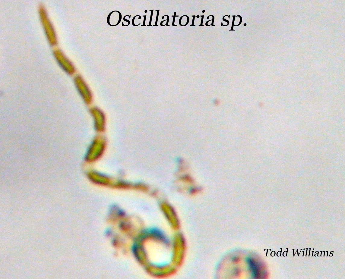
Example of Results: One of the organisms that I was never able to observe very well under the microscope was a Nematode sp. They were always moving and whipping around way to fast for me to over observe them. My process enabled me to view them for the first time, and I was even able to take a photo.


Sources: 
Conversation with Dr. McFarland on 11-16-2012
Photo: Nematode sp. by Todd Williams
Book: Pennack, Robert. " Nematode sp." Freshwater Invertabrates of the U.S. John Wiley and Sons.          Canada. p228


Week 5 - Final Observations

Hello, This is my last and final observation of my MicroAquarium. I hope you have enjoyed tagging along with me, as I observed my aquarium each week, and explored the world of micro-organisms.

First off, I observed my tank with my naked eye, I didn't notice any changes from last week. I could see a few Paramecium sp. but that is all I could see without using a microscope. Comparing my aquarium to the first week, I can say that some of major changes would be the sediment like I refered to last week. It is a lot more fuzzy than it was during week one. I asked Dr. McFarland if he thought it was waste from the living organism, and that was his best guess at what It could be. The water clarity, has stayed pretty much the same throughout these 5 weeks. The plant life I added, has turned yellow in some places. I believe it is on its last leg, and probably wouldn't last to many more weeks. 

Next, I observed my aquarium under a compound microscope. The first think I noticed, was that there were a large amount of diatoms, the population has grown tremendously from last week. I noted a few new organisms, that I had not seen in previous weeks. One, was a Halteria sp. According to the book: Free-living Freshwater Protozoa, it said that these are one of the most important herbivores in the aquatic ecosystem. They are number one, in the food chain, providing food for many of organisms. I have included a photograph of one below. They can be indentified by their circular shape, and flagella which radiate out like sun rays. I would say that the overall activity of organisms has decreased some this week. I would say that out of all five weeks, that week three had the most activity. Right around the time I added additional food. Although I would say that organisms that obtain food through photosynthesis, like Cynobacteria and Diatoms have increased in population. One type of cyanobacteria that I have seen a lot of is, Oscillatoria sp. According to the book: Freshwater Algae, by G. Prescott, they are one of the most common cyanobacteria, and are called Oscillatoria, because they slowly oscillate using small flagella. I have also included a photo of this species below. There was one organism, that I have seen quite frequently, but It has always been to quick to observe it under the microscope or take a photo of it. That organism is a Nematode sp. or commonly known as a roundworm. I have seen dozens of these in my aquarium over the past five weeks, which is kind of disconcerting thinking how many of these live in lakes and rivers that I may swim in.
by Todd Williams , 11-16-2012

by Todd Williams, 11-16-2012


There were a lot of organisms that looked very interesting, but moved to quickly for me to view under a microscope. This got me thinking, that it would be great if I could introduce a paralytic into the water to paralize the organisms, and make it easier to view them. This led me to conduct a quick experiment before I disposed of my aquarium at the end of my viewing session. On the next post I will tell you exactly what I did.

Sources:
-Conversation on 11/16/2012 with Dr. McFarland
-Patterson, D.J., "Halteria sp." Freeliving Freshwater Protazoa. New York. 2003
-Prescott, G,. "Oscillatoria sp." Freshwater Algae. McBrown Company. Iowa. 1996. p.123
-Photo of Halteria sp. by Todd Williams
-Photo of Oscillatoria sp. by Todd Williams

Saturday, November 10, 2012

Week 4 Observations

Hey Guys! I am back again, come check out what I discover this week as I look...

Just below the Surface!

For my first observation, I first used my naked eye to see what I could observe. Well, I do not see those white colored paramecium sp. That I saw last week moving around anymore. The tank looks pretty still. One thing that clearly came to my attention this week was that the sediment at the bottom of the tank, on week one it looked very thin, made up of clear sediments. Today it looks a lot thicker. It has almost doubled or tripled in thickness. It also looks very fuzzy. It has a much more grey color than the brownish color seen in the previous weeks. I am not quite sure what the reason for this is. Wait, I just saw one white paramecium, But other than that the tank looks still from the view of a naked eye.
Let’s take a look under the microscope. One comment I must make, I have just had a realization. In previous weeks, I was only mostly using the 10x objective on my compound microscope, and that has produced ample magnification to be able to see organism activity in my aquarium. This week, I am using the 40x objective most often. I at appears as if many of the organisms I have seen in previous weeks have died off, or are present in much fewer numbers. On the other hand, I have noticed a large population gain in many smaller organisms. Today,  I am going to talk a little more about some of the smaller organisms I am seeing today. I have seen many colonies of bacteria, all over the tank. They look like groups of tiny air bubbles. Most likely, they are living off of some particulate of dead organic matter that is present in the water. The organism that I have seen the most of today, has to be Diatoms. I have seen several  different shapes and sizes of them as well. According to the book, A Guide to Microlife by Kenneth Rainis and Bruce Russell, Diatoms are a major group of Algae. They are known for a unique cell wall, that is made of silica. Here are a few of the Diatoms that I observed today.
Fragellaria genus. (rectangular shaped) I have seen several of these in my aquarium. One has divided into two cells but has not yet made a ribbon. Each strip seen is a new cell.


Sources:
-Photo 1: Fragellaria genus. By Todd J Williams
-Photo 2: Cyclotella genus. By Todd J Williams
-Photo 3: Meridon genus. By Todd J Williams

-Rainis, Kenneth;  Russell,Bruce. A Guide to Microlife. Franklyn Watts Publishing, 1996. Connectcuit.   Diatoms pg. 113

-Canter-Lund, Hilda; Lund, John. Freshwater Algae. Biopress Limited, 1995. United kingdom 


Sunday, November 4, 2012

Week 3 Observations

Hey Guys,
So, My first observations this week were done with my naked eye. Using only my eye, I could definitely see what appeared to be tiny white worms moving slowly around my M.A. What was the most suprising was that they seemed to swim towards the top of my tank which is a definite change from past behavior I have observed. In the past it seems that most of the organisms have been down at the bottom of the tank, near the sediments that line the bottom of the tank. This observation made me question if some organisms were wandering away from the bottom of the tank in search of food.

I next used a compound microscope to view my aquarium. I was able to identify the white worms, I had seen earlier with my eye as a type of Paramecium sp. Under the microscope, these organisms are by far the largest living in my tank. They move quite slowly, in a worm-like lurching movement, which makes they pretty predictable and easy to observe under a microscope. The next significant thing I noticed was several dead organisms in my tank. With the help of Dr. McFarland, I was able to identify the exoskeleton of a dead Daphina sp., also known as a water flea. This brought my to think that some organisms, had probably died because of lack of food, and in turn became food for other organisms in the aquarium. It appears I have my vary own mini ecosystem living in my aquarium. I also noticed a lot more activity in my aquarium this week. In a conversation I had with Dr. McFarland, we discussed that the increase in activity was due to the fact that some organisms had died, and others were thriving off of their decaying bodies.
If no food were added, eventually all of the organisms would die off. So to prevent this "one"  pellet of beta fish food added to the tank on Friday, October 26, 2012. Here is some more precise information about what type of food I used, and the contents of the food. The brand was  "Atison's Betta Food" made by Ocean Nutrition, Aqua Pet Americas, 3528 West 500 South, Salt Lake City, UT 84104. The ingredients in the food were as follows: Fish meal, wheat flower, soy meal, krill meal, minerals, vitamins and preservatives. Analysis: Crude Protein 36%; Crude fat 4.5%; Crude Fiber 3.5%; Moisture 8% and Ash 15%. 
In the bottom of the tank, I believe I found where the pellet landed in the sediment. There were many organisms, many to fast to identify, near one spot at the bottom of my aquarium. I wonder if this food will cause the population of micro-organisms to increase in number to rapidly. I have a hypothesis that next week, when I observe my aquarium that there will be a lot more dead organisms, because I do not believe that there is enough food in the tank to support all the new life.
Here is a photo of the Exoskeleton of a Daphina sp. which I talked about earier in my blog. This was  one of several dead organisms I found in my tank this week, and a reason I added additional food.


Sources:
- Botany 111 Class Blog - Http://www.botany1112012.blogspot.com
- Dr. McFarland- Oral conversation on October 2, 2012 11:30am
- Atisons Betta Food made by Ocean Nutrition, Aqua Pet Americas Inc.
-Patterson, D.J., "Daphina sp." Freeliving Freshwater Protazoa. New York. 2003
-Photo: Exoskeleton of Daphina sp. by Todd Williams


Monday, October 29, 2012

Week 2 Observations

Hey Guys,
This week I must say, there seems to be a lot more going on activity wise in my aquarium. Lets take a look!
From the naked eye, I can see that the water level had dropped some, the water also appears to be a bit more cloudy compared to last week. It appears to have a slight brownish tint to it. I can see several organisms swimming around, which I could not see at all last week.
Ok, now using my microscope, I can definitely see there is a whole lot more activity. There are are a whole lot more organisms moving around compared to week one. Some seem to fly across the aperture at amazing speeds. Others, which I believe are some kind of Amoeba or protazoa, seem to lurch around like a slug.

This one species, which I have identified as a Cyclops sp. seems to have a flagella like tail that propels him through the water, in small bursts. I also identified a Paramecium sp., which looks a lot like a slug. It moves very slowly through the water and sifts through the sediment for food. I also identified, and took a great photo of a Urocentrum sp. This organism spins like a top in the water, around and around. According to the book "Free living Freshwater Protozoa" they use their tail to spin in that motion, and they feed off of bacteria in the water. I did notice that there was a lot more activity towards the bottom of the tank, but compared to week one, there was more activity in the top of the tank than the previous week. I have attached some photos I took with a camera attached to a microscope. I hope you enjoy! (Sources at bottom of blog.)


Sources:
-Patterson, D.J.,"Cyclops sp. Urocentrum sp., Paramecium sp." Freeliving Freshwater Protazoa. New York. 2003

-Photo: Cyclops sp., Utrocentrum sp., Paramecium sp. by Todd J. Williams


Monday, October 22, 2012

Week 1 Observation

A few mintutes after I set up my MicroAquarium, I placed it under a microscope and took a look at what was going on at a microscopic level. I observed some very fast organisms, that were moving to fast for me to identify them with my microscope. After moving my objective around the tank for a few mintutes, I settled in a focused at the bottom of the tank, where there was some sediments. I noticed a lot more activity down at the bottom, and I was able to focus in on a few organisms that were slow enough.
 I was able to identify a few Daphina sp. which is a microscopic organism that looks like a shrimp. According to the National Institute of Health website, a Daphina sp. is a micro-crustacean and is also referred to as a water flea. Next week I look forward to using a digital camera microscope and getting some good photos of what organisms I find in my tank. See you next week!
Sources:
-"Daphina" National Institute of Health. 2012 http://nih.gov/science/models/daphina/
- Daphina sp. Photo - Todd J. Williams (coming soon)

MicroAquarium™ Setup

On Wednesday October 17, 2012,
 I set up my MicroAquarium™ (M.A.). To find out more about exactly what a M.A. is, check the tab on the right side of my blog. To do this, the first thing I had to decided was what water source I was going to choose for my M.A. I was given the choice of choosing from Twelve different water sources. I chose my water source from the Fountain City duck pond. I chose this water source because I frequent the park around the pond, and wanted to see what microscopic life was taking place below the water. I posted some additional information on my water source on the right side of my blog page. In the next entry I will discuss what steps I took to set up my M.A.


After I had selected my water source, I used a water dropper to draw up water from a large bowl containing the pond water. I filled up the first 1/3 of my M.A. with water from the bottom of the bowl. I made sure to draw up a little mud and sediment with my water. I did this because many micro-organisms enjoy living in the sediment and mud at the bottom of a pond. Next, filled up the middle 1/3 with water from the middle of the bowl, and then the top 1/3 with water I skimmed off the surface of the water.
 I next inserted two species of aquatic plant material into my M.A. The plant I placed on the right was Amblystegium sp. This is a aquatic moss that was collected on 10/9/2011, from a natural spring at Carters Mill Park in Knox Co., TN. I have included further information about this plant and where it was collected in the tab on the right under the photo.  
The second plant, which I placed on the left side of my M.A., was Utricularia gibba. This is a flowering carnivous plant that was originally collected from Spain Lake, in White Co., TN. The collected species was then grown in water tanks, outside of the Hesler Biology Building at The University of Tennessee untila sample was collected for this experiment. I also have included further information about this plant and where it was collected in the tab on the right hand side of the page.

The  reason I added plant life to my M.A. was to oxygenate the water. Oxygen is essential for the micro-organisms living in the M.A. to survive.

(Sources at bottom of blog)